Tuesday, February 12, 2013

Ask Me


Feeding Tube Awareness Week might have ended yesterday but that doesn't mean we have to stop spreading awareness or knowledge. Feeding tubes are somewhat of an unknown to the majority of people. There are alot of missunderstandings and wrong information floating around out there and S4L wants to help fix that. Liam has been on a feeding tube for quiet some time now and has had 3 different types of tubes. Go ahead and ask any questions or state any concerns you have and we will be happy to answer. Comments are welcome. If you want to stay annonymous just email me at shootingforliam@yahoo.com You can also post questions at http://www.facebook.com/ShootingForLiam
 
Liam after Gtube and Nissin Fundo Surgery. October 2011

Here's some Questions we got on Liam's FB page:

Q: I have been following Liam's journey since my son was diagnosed with CDH inutero in Dec 2011. I have followed your ups and downs with Liam's feeding and am pretty worried we are gonne be on the same journey. We have a GJtube placed tomarrow at 9a instead of his Gtube. I would like any advice on how to care for a GJ that you could give me.

A: Feeding tubes can be over whelming at first. When Liam came out of surgery for a Gtube I was petrified to hold him because I thought Id hurt him. It took me weeks to realize I could hold him like a normal baby. And when it came time he did pull out the tube, he acted like nothing happened.

*As for care: keep the site cleam. Use a wet wash cloth to clean the area and keep it as dry as you can. Our medical supply CO provides us with split 2x2 guaze to use around the tube but I found that they don't work very well. They didn't soak up all the leaking fluid and the pad itself would be drenched in no time. The guaze had to be changed every few hours or they would just contribute to irritating the skin. I ordered a few Gtube pads a few weeks ago and within 2 days I saw a HUGE improvement. Check out Sew Amazing Creations. She makes really awesome tube pads at the cheapest price on the market. Only $1 per pad! http://www.facebook.com/SewAmazingCreations

*The most imortant thing with a GJtube is that you do NOT want it to spin. If it spins the J port could back out of the intestines into the stomach defeating the purpose of the GJ. Again the best thing I found to keep this from happening is the Gtube pads.

*Keeping your child from pulling out tube is a challenge but it is possible. We used to think a onsie would prevent Liam from pulling out his tube but last August we learned how very wrong we were. He was able to pull it out right with no problem whatsoever. Tummy belts work really well because the child cannot grab ahold of the tube with their hands and yank on it. I have this problem with Liam. There are many places online where you can order these but they are costly. Decorative yes but I know I cant afford $20 to $40 for one. Cherubim has a inexpensive tummy belt that works really well. Its stretchy and has built in velcro that makes it super easy to get on your child. They run $8. http://cherubim.storenvy.com  You can also use an ace bandage. The extensions are another issue. We use POSEY's (those things the hospital uses to strap on your little ones Pulse Ox) to strap the extensions to Liam's leg. It requies two now that he's bigger. When he was shorter and skinnier we were able to use one and strap them to his ankle. Works like a charm. We also thread his enstensions through his pant legs so it hides them from him.

*Pump feeding and mobility are a daily issue when you have an active one like Liam who just cant sit still. We figured out that we can put the pump and bag into its portable backpack and strap it to his walker allowing him to roam the house at his pleasure. Allowing him to free roam and crawl around without getting too far away from the pump is an issue. We have to just watch him and keep him near the pump or follow him around with the pump. As he's getting older we've noticed that he somtimes listens when we tell him to stop as he's crawling too far away but he also loves to ignore people. The other thing we've notices that when he's getting too far away and it pulls the tube just slightly he feels it and stops himself from going further away. He knows he doesnt like the pain. I think that as they get older it gets easier on everyone.

Q: My son has had a Gtube since July. We have had nothing but problems with it since his 6 week post op appointment. It has leaked, his skin is being eaten. They want to put in a GJ in cause he's throwing up past his Nissin and is failure to thrive.

A: Thats how Liam is. His tube leaks all the time. The Gtube pads are the best thing Ive found. They are thick so they pull the tube taunt against the inside preventing fluid from leaking through. Thats something the guaze doesn't do. We've tried all kinds of creams the GI prescribed but the best thing was to keep it dry.

Q: What problems have you had with the GJtube?

A: The main issue is that for some reason his GJtube actually desinigrates. The ports pop out because of this and his J port line has gotten clogged many times. We're not sure if its Liam or the way these tubes are made. Theres nothing we can do to prevent the tube system from falling apart but we can prevent it from getting clogged. Once a week we have to use soda, we use Pepsi but you can use Coke, to clean the Jport. We use 6 to 8 mls of soda, inject it into the J extension then let it set for atleast 15 to 20 minutes. Then we can go back to using it as normal. These are orders via our GI. Because its such a small amount it won't hurt him. And Ive monitored him after and theres no hyperness are anything else we've noticed after the soda gets into his system.


Liam's first feeding tube was an NG tube in the NICU. Because of the Cpap machine they have to put it in through the mouth. The second time he got his NGtube was in September of 2011.

GJtube Button


Sunday, February 10, 2013

Princess Alexandria Update

If you've all been keeping up on Facebook you know that Princess Alexandria got sick and ended up in the ICU on bipap. They said she'd be there for several weeks. She finally got out of ICU and onto the regular floor on just oxygen. Everyday she was getting better and stronger. Today mom posted that Alexandria might get released tomarrow. Make sure you don't say it outloud so that Alexandria cant hear you because these CDH babies have a communication line going. As soon as its said that they might go home, they tend to pull tricks that ends up in more time at "club med". We're praying for you Princess and can't wait to read the post that says your home <3 <3 <3

Feeding Tube Awareness Week


It's feeding tube awareness week again. Time to spread awareness as well as facts. Time to break the stereotypes.

What do I hate most about the feeding tube?
The stares I get from strangers when I take my son out in public while on his feed. He's only 18 months now but soon he will be old enough to recognize the looks of strangers and those looks are going to make him feel different, out of place, and maybe even like a "freak". As it is I get glares, questioning looks, and looks of distaste from strangers who see Liam on a feeding tube. No child, or parent should have to deal with this stress.

Commonly asked questions:

Why does your child need a feeding tube?
The requirement of feeding tubes can vary from a medical condition, failure to thrive or many other reasons. In Liam's case he was Failure To Thrive (FTT). He couldn't eat enough orally to keep up with the additional calories his body required to grow and thrive. He would get tired halfway through a feeding, be breathing fast and just fall asleep on me. This is the case for many infants with or even without other medical conditions.

How long will your child be on a feeding tube?
This is a question I get asked a lot. Its hard to say how long a child will require a feeding tube as it depends on the child. Every child is different and has different needs. In Liam's case it started out with the doctors thinking he would only need his feeding tube for a year. Then the time line changed to possibly age 2 when he got his Gtube. Now that he has a GJtube and has a oral aversion to food theyre thinking he will need it 4 more years. When I get asked this question I simply respond "He will need the feeding tube until he is able to eat by mouth and gain weight without using the tube." There is no set timeline as to how long a child will need a feeding tube.

How many types of feeding tubes are there?
NG
G
NJ
ND
GJ
J
TPN
(Click here to find out more about these)

The Feeding Tube Awareness Foundation offers a lot of information about feeding tubes and spreading awareness.

Lets use this week and spread awareness any way we can. Post on Facebook or Tumblr. Tweet about it, post pics on instagram about spreading awareness. Examples of hashtags you can use: #feedingtubeawareness #i<3atubie #tubielove

If you have any tips on how to prevent feeding tubes from being pulled out, how to keep an active child near their pump or any other tips please share with us. You can post in comments, email us your tips or post on our Facebook page.


NG (Naso Gastric): Runs from the nose to the stomach

Saturday, January 5, 2013

CDH Awareness Celebration Balloon Release

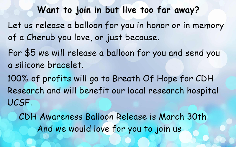
We're busy putting together our biggest event yet, the CDH Awareness Balloon Release as part of our celebration for CDH Awareness Week! Congress recognizes March 31st as CDH Awareness day, but this year Breath Of Hope is going for CDH Awareness WEEK! Yes you read that right, WEEK! And S4L is on board with this idea. On Saturday March 30th We will be doing a balloon release in Visalia CA in celebration of CDH Awareness week and to honor CDH survivors as well as CDH Angels. We are also selling CDH Awareness items to raise money for CDH Research. We have the date, March 30th, but we are still organizing the place and time.

 
And for our readers that live too far awar to attend or for whatever reason cannot attend, don't fret, we have an option that allows you to "participate":
 
 
We have CDH Awareness Items available for sale. They are available now as well as at the event.
 
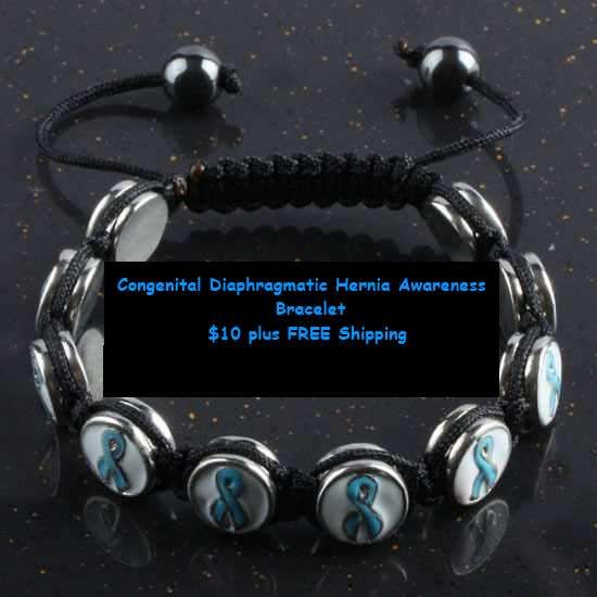
The teal CDH Awareness Macreme bracelet has been very popular and its hard to keep them in stock. They are $10 w/ free shipping. Because its so hard to keep them in stock it could take up to 4 weeks (sometimes longer) for you to get yours. We know the wait isnt ideal but we are very happy that so many of you have stuck with us even through the long wait time.
 
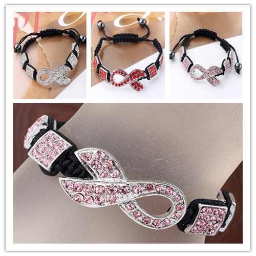
The Bling Awareness Bracelet has had many people interested even though I havent been able to offer teal for CDH Awareness. A few people order pink for their little ones currently fighting CDH or who have survived. This bracelet comes on Clear, Red and Pink only. Still trying to find a vendor that sells teal. They are $10 w/ free shipping and can also take a long period of time to come in as well.


CDH Awareness Teddy Bear's $8 plus Free shipping 5 1/2" tall
 
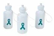
CDH Awareness Water Bottles $8 plus free shipping Holds 18 oz
 
 
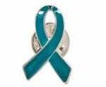
CDH Awarenes Teal Metal ribbon pins $5 plus free shipping
 
 
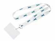
CDH Awareness Lanyards/badge holder $6 plus free shipping
 
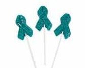
CDH Awareness Suckers 6 for $5 plus free shipping (online price only)
 
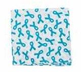
CDH Awareness Bandanas $4 plus free shipping
 
 
We also have Teal silicone bracelets $4 each or 4 for $12 plus free shipping.
 
If you are interested in ordering any of these items please email shootingforliam@yahoo.com so that we can send you a PayPal Invoice. Remember 40% of all sales go to CDH Research.


Saturday, December 29, 2012

RSV: Symptoms and prevention


RSV:
 Respiratory syncytial virus (RSV) is a very common virus that leads to mild, Respiratory syncytial virus (RSV)cold-like symptoms in adults and older healthy children. It can be more serious in young babies, especially to those in certain high-risk groups.

Children or babies with Congenital Diaphragmatic Hernia, Chronic Lung Disease and Severe Asthma are at high risk for RSV.

RSV is the most common germ that causes lung and airway infections in infants and young children.

Most infants have had this infection by age 2. Outbreaks of RSV infections most often begin in the fall and run into the spring.
The infection can occur in people of all ages. The virus spreads through tiny droplets that go into the air when a sick person blows their nose, coughs, or sneezes.
You can catch RSV if:
  • A person with RSV sneezes, coughs, or blows their nose near you
  • You touch, kiss, or shake hands with someone who is infected by the virus
  • You touch your nose, eyes, or mouth after you have touched something contamined by the virus, such as a toy or doorknob.
RSV often spreads very rapidly in crowded households and day care centers. The virus can live for a half an hour or more on hands. The virus can also live for up to 5 hours on countertops and for several hours on used tissues.
The following increase the risk for RSV:
  • Attending day care
  • Being near tobacco smoke
  • Having school-aged brothers or sisters
  • Living in crowded conditions
Symptoms vary and differ with age. They usually appear 4 - 6 days after coming in contact with the virus.
Older children usually have only mild, cold-like symptoms, such as cough, stuffy nose, or low-grade fever.
Infants under age 1 may have more severe symptoms and often have the most trouble breathing

In general, RSV symptoms include:
Many hospitals and clinics can rapidly test for RSV using a sample of fluid taken from the nose with a cotton swab. They can also suction the nose to get mucus from deaper in.

Antibiotics do not treat RSV.
Mild infections go away without treatment.
Infants and children with a severe RSV infection may be admitted to the hospital .
Treatment will include:
  • Oxygen
  • Moist (humidified) air
  • Fluids through a vein (by IV)
A breathing machine (ventilator) may be needed.

Rarely, RSV infection can cause death in infants. However, this is unlikely if the child is seen by a health care provider in the early stages of the disease .
More severe RSV disease may occur in the following infants:
  • Premature infants
  • Infants with chronic lung disease
  • Infants whose immune system does not work well
  • Infants with certain forms of heart disease
In young children, RSV can cause:
Children who have had RSV bronchiolitis may be more likely to develop asthma.

Call your health care provider if breathing difficulties or other symptoms of this disorder appear. Any breathing difficulties in an infant are an emergency. Seek medical attention right away.

A simple way to help prevent RSV infection is to wash your hands often, especially before touching your baby. It is important to make certain that other people, especially caregivers, take steps to avoid giving RSV to your baby.
The following simple steps can help protect your baby from getting sick:
  • Insist that others wash their hands with warm water and soap before touching your baby.
  • Have others avoid contact with the baby if they have a cold or fever. If necessary, have them wear a mask.
  • Be aware that kissing the baby can spread RSV infection.
  • Try to keep young children away from your baby. RSV is very common among young children and easily spreads from child to child.
  • Do not smoke inside your house, car, or anywhere near your baby. Exposure to tobacco smoke increases the risk of RSV illness.
Parents of high-risk young infants should avoid crowds during outbreaks of RSV. Moderate-to-large outbreaks are often reported in the local news and newspapers to provide parents with an opportunity to avoid exposure.
The drug Synagis (palivizumab) is approved for the prevention of RSV disease in children younger than 24 months who are at high risk for serious RSV disease. Ask your doctor if your child should receive this medicine.

Synagis HELPS prevent RSV but its not 100%.
Washing your hands
Using hand sanatizer
Wiping down your countertops and other surfaces with Lysol wipes
Using Lysol Nuetra air to kill air born germs
Keeping your child out of stores during RSV, cold and flu season
Keeping sick visitors away
Wearing a mask when sick
All these things helps increase the chance of preventing RSV for your family.

I hear alot of people with stuffy noses or coughs say "Oh its just allergies". 95% of the time it turns out NOT to be just allergies, in my experience anyway. Don't take their word for it. Refuse their company or make them wear a mask and wash their hands. If your baby gets sick, theyre the ones suffering not you. Even if your child gets a cough and runny nose, remember RSV starts out the same way. You can take your child to your pediatrician to be checked. Its better to be safe than sorry. We wish you all the luck and pray you avoid a RSV breakout in your home.
 

Thursday, December 27, 2012

Feeding Tubes: Whats the difference?

Feeding Tubes: What's the difference?

A feeding tube is a medical device used to provide nutrition to patients who cannot obtain nutrition by mouth, are unable to swallow safely, or need nutritional supplementation. The state of being fed by a feeding tube is called gavage, enteral feeding or tube feeding. Placement may be temporary for the treatment of acute conditions or lifelong in the case of chronic disabilities. A variety of feeding tubes are used in medical practice. They are usually made of polyurethane or silicone. The diameter of a feeding tube is measured in French units (each French unit equals 0.33 millimeters). They are classified by site of insertion and intended use.

Conditions requiring tube feeding:
The most common types of tubes include those placed through the nose, including Nasogastric, Nasoduodenal, and Nasojejunal tubes, and those placed directly into the abdomen, such as a Gastrostomy, Gastrojejunostomy, or Jejunostomy feeding tube

Nasogastric Feeding Tube
(Sometimes NG tubes are placed through the mouth in cases like here when Liam was on CPAP. The NGtube is the orange tube going into his mouth)

 
A nasogastric feeding tube or NG-tube is passed through the nares (nostril), down the esophagus and into the stomach. This type of feeding tube is generally used for short term feeding, usually less than a month, though some infants and children may use an NG-tube longterm. Individuals who need tube feeding for a longer period of time are typically transitioned to a more permanent gastric feeding tube. The primary advantage of the NG-tube is that it is temporary and relatively non-invasive to place, meaning it can be removed or replaced at any time without surgery. NG-tubes can have complications, particularly related to accidental removal of the tube and nasal irritation.

Nasojejunal and Nasoduodenal Feeding Tube


A Nasojejunal or NJ-tube is similar to an NG-tube except that it is threaded through the stomach and into the jejunum, the middle section of the small intestine. In some cases, a nasoduodenal or ND-tube may be placed into the duodenum, the first part of the small intestine. These types of tubes are used for individuals who are unable to tolerate feeding into the stomach, due to dysfunction of the stomach, impaired gastric motility, severe reflux or vomiting. These types of tubes must be placed in a hospital setting.


Gastrostomy or Gastric feeding tube


A gastric feeding tube (G-tube or "button") is a tube inserted through a small incision in the abdomen into the stomach and is used for long-term enteral nutrition. One type is the percutaneous endoscopic gastrostomy (PEG) tube which is placed endoscopically. The position of the endoscope can be visualized on the outside of the patient's abdomen because it contains a powerful light source. A needle is inserted through the abdomen, visualized within the stomach by the endoscope, and a suture passed through the needle is grasped by the endoscope and pulled up through the esophagus. The suture is then tied to the end of the PEG tube that will be external, and pulled back down through the esophagus, stomach, and out through the abdominal wall. The insertion takes about 20 minutes. The tube is kept within the stomach either by a balloon on its tip (which can be deflated) or by a retention dome which is wider than the tract of the tube. G-tubes may also be placed surgically, using either an open or laparoscopic technique.
Some individuals continue to use a long, catheter-like tube, while others use a small "button" with a detachable extension set for feedings. Most G-tubes can be changed easily at home. Gastric feeding tubes are suitable for long-term use, though they sometimes need to be replaced if used long term. The G-tube can be useful where there is difficulty with swallowing because of neurologic or anatomic disorders (stroke, esophageal atresia, tracheoesophageal fistula), and to avoid the risk of aspiration pneumonia. However, in patients with advanced dementia or adult failure to thrive it does not decrease the risk of pneumonia.
What we like about the Gtube: If it gets pulled out you just put another in. No hospital visits required and no surgery.

Gastric drainage tube

A G-tube may instead be used for gastric drainage as a longer term solution to the condition where blockage in the upper reaches of the small intestine causes bile and acid to accumulate in the stomach, typically leading to periodic vomiting. Where such conditions are only short term, as in a hospital setting, a nasal tube connected to suction is usually used. A blockage lower in the intestinal tract may be addressed with a surgical procedure known as a colostomy, and either type of blockage may be corrected with a bowel resection under appropriate circumstances. If such correction is not possible or practical, nutrition may be supplied by parenteral nutrition.


Gastrojejunostomy feeding tube
(What Liam currently has)

A gastrojejunostomy or GJ feeding tube is a combination device that includes access to both the stomach and the jejunum, or middle part of the small intestine. Typical tubes are placed in a G-tube site or stoma, with a narrower long tube continuing through the stomach and into the small intestine. The GJ-tube is used widely in individuals with severe gastric motility, high risk of aspiration, or an inability to feed into the stomach. It allows the stomach to be continually vented or drained while simultaneously feeding into the small intestine. GJ-tubes are typically placed by an Interventional Radiologist in a hospital setting. The primary complication of GJ-tubes is migration of the long portion of the tube out of the intestine and back into the stomach.
What we like about the GJtube: Feeds go in through one port (J) leaving the stomach port (G) free for meds and to be vented. For Liam is very important that he has this port so that when he is sick and vommits we can vent him and "avoid" aspiration. This help prevent Liam from aspirating and it turning into Pnumonia. Its not fool proof but it helps.

Jejunostomy feeding tube

A jejunostomy feeding tube (J-tube) is a tube surgically inserted through the abdomen and into the jejunum (the second part of the small intestine). The procedure is called a jejunostomy. There are several techniques for placement, including a direct surgical or endoscopic technique, or a more complicated Roux-en-Y procedure. The J-tube may use a long, catheter-like tube or a button. Depending on the placement type, the tube may be changed at home, or may need to be changed at a hospital. A J-tube is helpful for individuals with poor gastric motility, chronic vomiting, or at high risk for aspiration.

The effectiveness of feeding tubes varies greatly depending on what condition they are used to treat.

Children

Feeding tubes are used widely in children with excellent success for a wide variety of conditions. Some children use them temporarily until they are able to eat on their own, while other children require them longterm. Some children only use feeding tubes to supplement their oral diet, while others rely on them exclusively.

Advanced dementia and adult failure to thrive

There is strong evidence that feeding tubes do not help patients with advanced dementia or adult failure to thrive, and expert opinion[2][3][4][5] recommends they not be offered to these patients. Studies have definitively proven to they do not prolong life, they do not decrease the risk of pneumonia, they do not improve wound healing, they do not help weight gain, and they do not help the patient regain any strength or functional ability such as walking or self-care. Patients with advanced dementia also often pull at their G tubes causing them to be dislodged, and frequently require physical restraints, such as tying their wrists to the bed, to keep this from happening.[6]

Eating disorders

Patients with the eating disorder anorexia nervosa may be tube fed if they are significantly malnourished. This can be voluntary or in some cases where the patient is resistant to feeding under the force of the Mental Health Act. Patients may tamper with their feeds, which can interfere with the effectiveness of feeding.

ICU

Nasogastric tubes are often used in the intensive care unit (ICU) to provide nutrition to critically ill patients while their medical conditions are addressed. There is moderate evidence for use of feeding tubes in the ICU, especially if requiring mechanical ventilation for more than three days.

Neurologic disease and mechanical obstruction

There is at least moderate evidence for feeding tubes improving outcomes for chronic malnutrition in patients with cancers of the head and neck, acute stroke while the patient undergoes rehab, and ALS.

Complications
Gastric feeding tubes have a variety of complications. As gastric feeding tubes are placed as part of a procedure that punches a hole in the stomach and skin, this can lead to leaking of contents into the abdomen causing severe infection and death. The most frequent complication is irritation around the site of the insertion, generally caused by stomach acid and feedings leaking around the site. Barrier creams, dressings, and frequent cleaning is generally recommended.[citation needed]
Nasogastric feeding tubes, if inserted incorrectly, can cause collapsed lungs and consequently, death; however this is an extremely rare complication.[7]
NG complications include the tube migrating up out of the stomach and into the lungs therefore "drowning" the patient. Also inserting the tube too far in or not far enough.


(info from http://en.wikipedia.org/wiki/Gastric_feeding_tube#Gastric_feeding_tube)


Mediport Information


Our little Liam has spent so much time in the hospital and has been poked so many times for IV's and blood draws that his veins blow. Blood draws are extremely difficult and IV's are near impossible and last usually no longer than 2 to 24 hours. PICC lines are a no go. He has so much scar tissue that they can only get a newborn sized line in him and cant use it to draw blood or give proper amount of fluids or medicine through it. His last hospital stay he went through 3 IV's in 2 days. One IV only lasted 2 hours before it infiltrated and left Liams arm swollen and painful.

(Infiltrated IV)
 
Because of his frequent stays at the hospital and all his other IV issues, he became a candidate for a mediport.
 
 
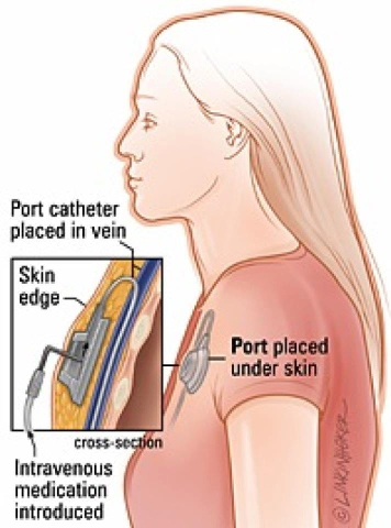
In medicine, a port (or portacath) is a small medical appliance that is installed beneath the skin. A catheter connects the port to a vein. Under the skin, the port has a septum through which drugs can be injected and blood samples can be drawn many times, usually with less discomfort for the patient than a more typical "needle stick".
Ports are used mostly to treat hematology and oncology patients, but recently ports have been adapted also for hemodialysis patients.
The port is usually inserted in the upper chest, just below the clavicle or collar bone, leaving the patient's hands free.

(Metal version on the mediport with needle attached)


How it works:
A port consists of a reservoir compartment (the portal) that has a silicone bubble for needle insertion (the septum), with an attached plastic tube (the catheter). The device is surgically inserted under the skin in the upper chest or in the arm and appears as a bump under the skin. It requires no special maintenance and is completely internal so swimming and bathing are not a problem. The catheter runs from the portal and is surgically inserted into a vein (usually the jugular vein, subclavian vein, or superior vena cava). Ideally, the catheter terminates in the superior vena cava, just upstream of the right atrium. This position allows infused agents to be spread throughout the body quickly and efficiently.
The septum is made of a special self-sealing silicone rubber; it can be punctured hundreds of times before it weakens significantly. To administer treatment or to withdraw blood, a health professional will first locate the port and disinfect the area. Then he or she will access the port by puncturing the overlying skin with a 90° Huber point needle although a winged needle may also be used. (Due to its design, there is a very low infection risk, as the breach of skin integrity is never larger than the caliber of the needle. This gives it an advantage over indwelling lines such as the Hickman line.) Negative pressure is created to withdraw blood into the vacuumized needle, to check for blood return and see if the port is functioning normally. Next, the port will be flushed with a saline solution. Then, treatment will begin. After each use, a heparin lock is made by injecting a small amount of heparinized saline (an anticoagulant) into the device. This prevents development of clots within the port or catheter. In some catheter designs where there is a self-sealing valve at the far end, the system is locked with just saline. The port can be left accessed for as long as required. The port is covered in a dressing to protect the site from infection and to secure the needle in position.
If a port is used infrequently, it may be necessary to access the port, flush it with saline, and inject a new heparin lock every month to prevent clotting between uses.
The surgery itself is considered minor, and is typically performed under both local anaesthesia and conscious sedation. Patients sometimes have a little discomfort after the procedure, and can be managed with a narcotic, such as demerol for 24–48 hours.
A port is most commonly inserted as a day surgery procedure in a hospital or clinic by a surgeon or an interventional radiologist, under conscious sedation. When no longer needed, the port can be removed in the operating room.

(xray of a mediport)


Uses:
  • To deliver total parenteral nutrition in those unable to take (adequate) food orally for a long period of time.
  • To deliver chemotherapy to cancer patients who must undergo treatment frequently. Chemotherapy is often toxic, and can damage skin and muscle tissue, and therefore should not be delivered through these tissues. Portacaths provide a solution, delivering drugs quickly and efficiently through the entire body via the circulatory system.
  • To deliver coagulation factors in patients with severe hemophilia.
  • To withdraw (and/or return) blood to the body in patients who require frequent blood tests, and in hemodialysis patients.
  • To deliver antibiotics to patients requiring them for a long time or frequently, such as those with cystic fibrosis and bronchiectasis.
  • Delivering medications to patients with immune disorders.
  • For treating alpha 1-antitrypsin deficiency with replacement therapy
  • For delivering radiopaque contrast agents, which enhance contrast in CT imaging.
  • To fill or withdraw fluid from the Lap-Band or Realize gastric bands used in Bariatric surgeries.
  • For paitents like Liam who are in the hospital alot and veins wont handle IV's or PICC lines

  • There are many different types of portacaths. The particular type selected is based on the patient's specific medical conditions.
    Portals:
    • can be made of plastic, stainless steel, or titanium
    • can be single chamber or dual chamber
    • vary in height (although the base is about the size of a quarter)
    Catheters:
    • can be made of biocompatible, medical-grade polyurethane or silicone
    • can vary in length (cm)
    • can vary in diameter (mm)
    Portacaths can be put in the upper chest or arm. The exact positioning itself is variable as it can be inserted to avoid visibility when wearing low cut shirts, and to avoid excess contact due to a backpack or bra strap. The most common placement is on the upper right portion of the chest, with the catheter itself looping through the right subclavian vein down towards the patient's heart. It can also be situated on the muscle that sits on the ribs with the tube coming up towards the heart.

    Risks:
    1. Infection - a severe bacterial infection can compromise the device, require its surgical removal, and seriously jeopardize the health of the already weak patient.
    2. Thrombosis - formation of a blood clot in the catheter may block the device irrevocably. To prevent clotting the portacath is flushed with saline and heparin, usually by a nurse or other medical professional, or someone properly trained that is a family member or the patient, at least once every four weeks, or more often in conjunction with administering medication.
    3. Mechanical failure - is extremely unlikely. It is possible in a rare event that part of the system could break, usually the attached catheter, and become lodged in the circulatory system. In such a case it is unlikely to cause serious harm; many patients are asymptomatic but the mechanical failure is discovered because of an inability to flush or withdraw fluids from the port. In those rare instances intervention surgery is required to withdraw the failed vascular access device.
    4. Age - If the device is put into a child, the child's growth means that the catheter becomes relatively shorter and will move away from the inferior part of the superior vena cava - it might be necessary to remove or replace it.
    5. Pneumothorax - Attempts to gain access to the subclavian vein or jugular vein can injure the lung, possibly leading to this complication. If the pneumothorax is large enough, a chest tube might need to be placed. In experienced hands, the incidence of this complication is about 1%.
    6. Arterial injury - The subclavian artery can be inadvertently punctured. This usually leads to a subcutaneous hematoma and occasionally a pseudoaneurysm. An alternative site may need to be used for port placement. Puncture of the carotid artery is more rare, since attempts to access the nearby jugular vein are increasingly done with ultrasound guidance.


    (Info gained from http://en.wikipedia.org/wiki/Port_(medical)